MRI
GENERAL OVERVIEW ABOUT THE EXAM
Magnetic Resonance Imaging, better known as MRI, is a safe, non-invasive imaging technique designed to generate detailed images of your body. Using a strong magnetic field, radio waves, and advanced computing, it generates cross-sectional “slices” of your body for in-depth examination, without any ionizing radiation.
MRI is a sophisticated imaging method, producing high-definition images of the head, neck, spine, muscles, joints, bones, and even the chest, abdomen, and pelvis. Occasionally, we might use a contrast agent, such as gadolinium, to improve image clarity and aid accurate diagnosis.
MRI's high-resolution imaging can detect minute changes in your body's structures, ensuring the most accurate diagnosis and best treatment plan. Despite its technical nature, rest assured, our team is here to guide you through every step, making your MRI experience as comfortable as possible.
Quick Links & Info

Scheduling
For information about your appointment or to schedule a new one call (386) 274-6000.

Exam Preps
Be ready for your next appointment.

Our Providers
See a full listing of our providers and their bios.
Types of MRI
RA offers MRI at our our St. Augustine, Palm Coast, Daytona Beach and Port Orange locations. We offer open-bore MRI, which is designed for younger, larger or claustrophobic patients at our St. Augustine, Palm Coast, Daytona Beach and Port Orange locations.
-
Musculokeletal MRI
-
Breast MRI
-
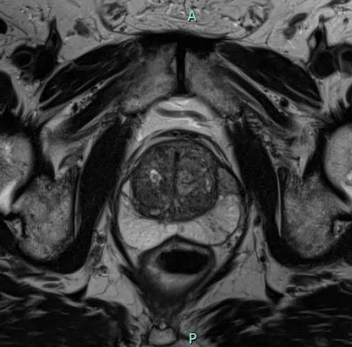
Prostate MRI
-
Cardiac MRI
-
Small Bowel MRI (MR Enterography)
-
MR Angiography
-
MRI Elastography - Liver
What to Expect
A contrast material may be injected or swallowed, depending on your exam. Because MRI utilizes magnets, you will be asked to remove all metal objects like jewelry, belts, etc. Most people with permanent metal situated anywhere in the head or body are not eligible for MRI examination. You will be asked to lie still on the table with the area being investigated situated inside the MRI machine while it captures a series of images. Earplugs will be provided to diminish the knocking and thumping sounds created by the scan. The test is painless, but can take from 30 minutes to 2 hours. Some people feel confined in an MRI machine. For people prone to claustrophobia, RA offers open-bore MRI, which offers a wider environment suitable for such patients. Open-bore MRI may also be indicated for people with larger bodies and children, who typically find open MRI more comfortable.

ACR Accredited in MRI
Our facilities have been accredited by the American College of Radiology (ACR) for accuracy, safety and best practice standards in MRI. For a complete listing of our accreditations per imaging center, please visit our Contact page.
Featured