Services

CT

GENERAL OVERVIEW ABOUT THE EXAM

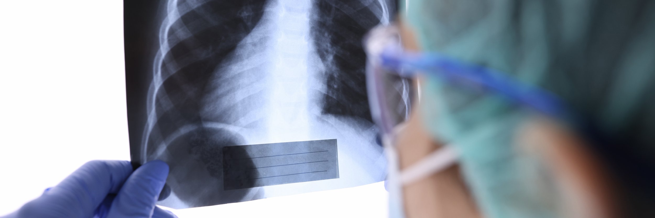
Computed Tomography, also called a CAT scan or CT, is a non-invasive medical test that uses a series of cross-sectional images to view a bodily organ, structure or system. The CT scanner consists of a table, which supports the patient, and a large ring that rotates 360 degrees around the area being studied, taking multiple images from every angle in just seconds. The images are sent to a computer screen to create a 3D composite that is sent to your doctor for review so that he or she can collaborate with your RA radiologist to form a diagnosis.


How a Low Dose CT Scan Could Save Your Life

Download the LDCT Lung Cancer Screening Brochure

CT IV Contrast Alert

Due to a national shortage of CT IV Contrast, you may receive a call from Radiology Associates to modify CT protocols.

We appreciate your assistance in this matter.


Types of CT

CT of the Body

CT scanning of the body is a process in which the scanner rotates 360 degrees around the body, capturing multiple cross-sectional images of bones, organs, soft tissues and blood vessels that are sent to a computer for reformation into highly-detailed 3D images on the screen, so your doctor and radiologist can rotate the image for views from every angle. The composite image can also be printed using a 3D printer, creating a model of the area being scanned. A CT body scan is used to locate, diagnose and stage treatment for a variety of diseases and disorders, including cancer and lymphoma, infection, heart and cardiovascular disease, internal injury or trauma, musculoskeletal problems and many others.

CT produces clearer images than traditional x-ray, and produces exceptionally fast imaging results of nearly every type of organ, tissue and vessel in the body, making it one of the most effective tests for detecting cancer, lymphoma and tumors in the chest and lungs, abdomen and pelvis, liver, kidney, pancreas, ovaries and prostate. CT’s 3D imaging capacity not only finds tumors, it actually offers information about their size, number, location, shape and invasion into adjacent tissues and organs.

CT is highly useful in early detection of vascular diseases such as blockage, vessel narrowing, aortic aneurysms and clots, problem that, when caught and treated early, can help prevent stroke, heart attack, kidney failure and other potentially mortal conditions.

Low Dose Lung Cancer Screening CT

Low Dose Computed Tomography (LDCT) is a reduced-radiation chest screening designed to produce very detailed images and promote the early discovery of nodules and abnormalities in the lungs. This quick, painless, non-invasive test captures multiple images of the lungs that are sent to a computer for a 3D view your radiologist can use to identify cancer before it causes symptoms, like coughing, wheezing, shortness of breath, which usually indicate cancer has spread outside the lungs, where it is often untreatable.

People ages 50 and older with a 20 “pack-year” history (1 pack a day x 20 years, 2 packs a day x 10 years, etc.) who currently smoke or have quit within the past 15 years, and people 55 and older with 30 pack years and a personal or immediate family history of lung/respiratory disease, may be candidates for LDCT lung cancer screening.

LDCT is a screening test for people without symptoms and requires a doctor’s referral. If you’re between 50-80 years of age with a 20-30 pack-year history and a physician’s referral, your scan will likely be covered by Medicare. If you are or were a heavy smoker, talk to your doctor about whether LDCT might be for you.

CT Colonography

Colon cancer is unfortunately one of the most commonly diagnosed cancers in the United States and is also the second leading cause of cancer-related deaths.

Colon cancer screening is currently recommended beginning at age 50 for average risk patients. If you are at higher than average risk, screening may need to begin before age 50.

The traditional method for colon and rectal cancer screening is with optical colonoscopy. In this procedure, the patient is sedated and a small, flexible, catheter-type device with a camera is inserted into the colon through the rectum.

RA offers CT Colonography (also known as Virtual Colonoscopy) as a method to screen for colorectal cancer. This method is less invasive and time-consuming than traditional optical colonoscopy with no needles or sedation required. This examination is performed on our CT scanners after insufflating the colon with air. Once the examination is complete, the imaging data is evaluated by one of our radiologists on a special computer workstation.

Small Bowel Imaging (CT Enterography)

This imaging study is a newer alternative to traditional barium fluoroscopy exams to evaluate the small bowel while using a multi-detector CT scanner. The most common indications include assessment for inflammatory bowel disease (Crohn’s disease), gastrointestinal bleeding, and small bowel tumors. This technique is also very helpful in differentiating between new (active) and old (fibrotic) strictures in patients with Crohn’s disease. Differentiating active versus fibrotic strictures has important implications regarding whether a patient receives medical management or surgery.

What to Expect

Upon arrival, you may be asked to change from your regular clothes into a gown that has no metal zippers or snaps.  A locker will be provided for your clothes.

If your medical provider has ordered an exam with intravenous contrast, an IV (intravenous) line will be placed in your arm. If an abdomen and/or pelvis CT has been ordered, you also may have to drink oral contrast material depending on the clinical indication. Oral and intravenous contrast can be extremely helpful (and sometimes mandatory) to appropriately diagnose or exclude certain medical conditions. RA’s care team reviews all orders to ensure that you receive contrast agents only when necessary.

During your CT examination, you will lie on a table that will slide through an open circular or donut-shaped structure.  For many exams, you will be asked to hold your breath during image acquisition.  Once acquired, the image data will be reconstructed by the computers and the final exam will be reviewed by one of our radiologists. A detailed report will be sent to your health care provider.

For Small Bowel CT Imaging:

The exam will be performed on one of our multi-detector CT scanners following a few special steps to prepare the small bowel. You will be asked to have nothing by mouth (NPO) for at least 4 hours prior to the exam. This ensures that any ingested food material is not confused for an abnormality. You will also need to arrive at our imaging center about one hour prior to the examination time in order to drink a special oral contrast agent to distend the small bowel. Once these steps are complete, you will be placed on the CT scanner and images will be obtained rapidly following the injection of contrast material.


ACR Accredited in CT

Our facilities have been accredited by the American College of Radiology (ACR) for accuracy, safety and best practice standards in Computed Tomography (CT). For a complete listing of our accreditations per imaging center, please visit our Contact page.